Recombinant Antibodies
- ‹ Primary Antibodies
- Guide to Primary Antibody Types
- Recombinant Antibodies
- Monoclonal Antibodies
- Recombinant Rabbit Monoclonal Antibodies
- Polyclonal Antibodies
- Conjugated Primary Antibodies
- Control Antibodies
- Epitope Tag Antibodies and Related Antibodies
- Research Area Antibodies
- Signal Pathway Antibodies
- Cell Marker Antibodies
- Rabbit Monoclonal Antibodies
Invitrogen recombinant rabbit monoclonal antibodies are derived from rabbit monoclonal antibody-producing cell lines. These cell lines are developed by isolating and cloning the specific antibody heavy and light chain DNA sequences. These recombinant cloned antibodies are not susceptible to cell-line drift or lot-to-lot variation, allowing for peak specificity and performance.
Recombinant rabbit monoclonal antibody production
Recombinant rabbit monoclonal antibodies are similar in consistency to traditional mouse monoclonal antibodies while offering an animal-origin free option. Rabbits are immunized and, instead of using hybridomas, peripheral blood mononuclear cells (PBMCs) are isolated and screened. After the DNA is cloned as a library and screened, it is subcloned to a single clone and screened again. The resulting DNA is used for production of animal origin-free formulations by cell culture.
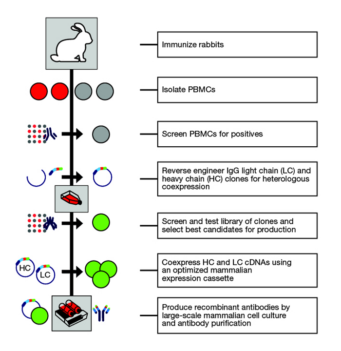
Figure 1. Recombinant rabbit monoclonal antibody production.
Advantages of recombinant rabbit monoclonal antibodies
Recombinant rabbit monoclonal antibodies exhibit superior lot-to-lot reproducibility because banked DNA does not exhibit cell drift – a phenomenon where hybridomas change expression patterns over time. Because the screening processes used in making recombinant rabbit monoclonal antibodies involves three different screening steps, the resulting antibody has better specificity and sensitivity than standard antibodies.
Other advantages include:- Better specificity and sensitivity compared to standard antibodies
- Lot-to-lot consistency due to recombinant technology
- Animal origin-free formulations, the result of production by cell culture
- Improved antibody performance due to multiple screens during development
Improved antibody performance due to multiple screens during development
Need a custom recombinant rabbit monoclonal antibody? Request a quote >
Reproducible manufacturing process
Recombinant rabbit monoclonal antibodies are manufactured by transfecting mammalian cells with heavy and light chain antibody cDNA. This highly reproducible process results in unparalleled lot-to-lot consistency. This consistency saves time and money because experimental conditions do not require revalidation. Figure 1 shows the consistent western blotting results achieved using independent lots of a recombinant rabbit monoclonal antibody, and its graphical representation. Figure 2 shows the lot-to-lot consistency in immunocytochemistry, and its graphical representation. Figure 3 shows the same results in flow cytometry.
Figure 3 shows the same results in flow cytometry.
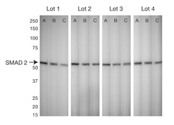
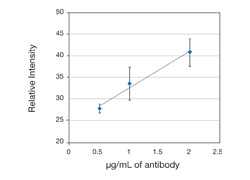
Figure 1. Lot-to-lot consistency using western blot.HeLa cell extracts separated on reducing gels were probed with four different lots of SMAD2 recombinant rabbit monoclonal antibodyThe concentrations in lanes A, B, and C were 2 μg/mL, 1 μg/mL, and 0.5 μg/ mL, respectively. Primary antibody was detected using WesternBreeze Chemiluminescent Kit–Anti-Rabbit.Left is quantitative representation of four lots at different concentrations, showing best lot-to-lot consistency (n=4, Error bars=standard error).
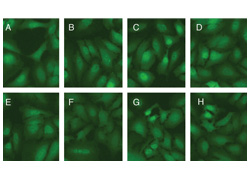
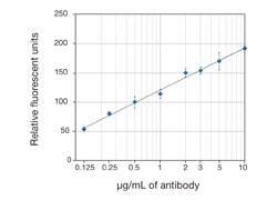
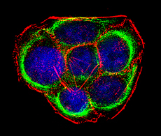
Figure 2. Lot-to-lot consistency using immunocytochemistry.Tissue cultured HeLa cells stained with four different lots of SMAD2 recombinant rabbit monoclonalantibody.A through D represents Lots 1 through 4 at concentration of 3 µg/mL, E through H represents Lots 1 through 4, at concentration of 0.5 μg/mL. Alexa Fluor 488 goat anti-rabbit IgG at 1:1000 was used as secondary antibody. Left is quantitative representation, of four lots at different concentrations, showing best lot-to-lot consistency (n=4, Error bars=standard error).
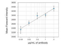
Figure 3. Lot-to-lot consistency using flow cytometry. Quantitative representation of the flow cytometry, with four independent lots of SMAD2 recombinant rabbit monoclonal antibodyat various concentrations, showing high lot-to-lot consistency. HeLa cells were fixed and permeabilized using FIX & PERM Reagents.SMAD2 ABfinity Antibody incubation was followed by Alexa Fluor 488 goat anti-rabbit IgG(n=4, Error Bars= Standard Error).
Reliable sensitivity and specificity
Recombinant rabbit monoclonal antibodies are more sensitive and specific than other antibody formats. These antibodies only react with the target of choice, eliminating detection of the wrong signal due to unspecific binding. More highly sensitive antibodies can detect very low-level targets that may be difficult to detect with other antibodies. Plus, precious samples are saved, needing less antibody for detection.
Figure 4 shows a direct comparison of a recombinant rabbit monoclonal STAT4 antibody with the best commercial STAT4 antibodies in western blotting. This comparison includes antibodies from polyclonal, traditional hybridoma monoclonal, and rabbit hybridoma monoclonal platforms. Figure 5 demonstrates the same results in immunocytochemistry.
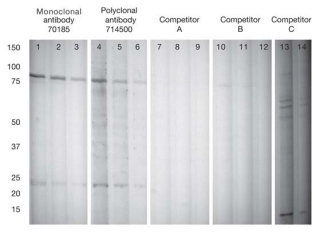
Figure 4. Superior western blotting results obtained using recombinant rabbit monoclonal antibody.The STAT4 recombinant rabbit monoclonal and polyclonal antibodies were used at 2, 1, and 0.5 μg/mL. STAT4 antibodies from other vendors were used at their recommended concentrations. Primary antibody was detected using WesternBreeze Chemiluminescent Kit–anti-Rabbit.
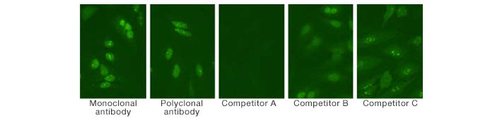
Figure 5. Enhanced immunocytochemistry results obtained using recombinant rabbit monoclonal antibody. Immunocytochemistry results while using STAT4 recombinant rabbit monoclonal antibodyat 2.5 μg/mL compared with the same antibodies as in Figure 4. STAT4 antibodies from other vendors were used at their recommended concentrations. Alexa Fluor 488 goat anti-rabbit IgGat 1:1000 was used as secondary antibody.
Antibody validation
Our recombinant rabbit monoclonal antibodies are validated and characterized by multiple applications. This extensive validation process allows confidence in target specificity, without any need for optimization. Examples of their application are shown in Figures 6–9.
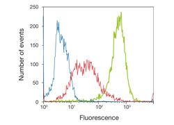
Figure 6. Flow cytometry of Jurkat cells labeled withAKT [pS473] - Recombinant Rabbit Monoclonal Antibody.Jurkat cells were incubated with 50 µM PI3K/AKT signaling pathway inhibitor LY294002 (red trace) or without (green trace) for 1 h prior to being fixed and permeabilized using FIX & PERM reagentsCells were then stained with 1 μg/test of Recombinant Rabbit Monoclonal Antibodyfollowed by Alexa Fluor 488 goat anti-rabbit IgG.The blue trace represents secondary antibody alone.
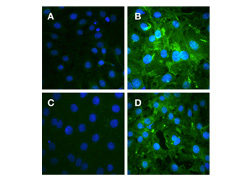
Figure 7. Immunocytochemistry of mouse fibroblasts cells labeled withAKT [pS473] - Recombinant Rabbit Monoclonal Antibody. Mouse fibroblast cells were treated with (A) or without (B) 10 µg/mL insulin and labeled with rabbit anti-AKT [pS473] (5 µg/mL). Signal is knocked down after incubation with the phosphopeptide used as antibody immunogen (C) but not with the non-phosphopeptide (D). Alexa Fluor 488 goat anti-rabbit IgG at 1:1000 was used as secondary antibody, nuclei are stained with Hoechst (blue).
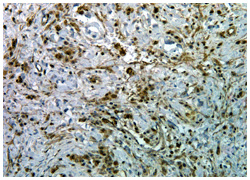
Figure 8. Immunohistochemistry of human esophagus carcinoma tissue labeled with AKT [pS473] - Recombinant Rabbit Monoclonal Antibody.Formaldehyde-fixed paraffin-embedded (FFPE) human esophagus carcinoma tissue was labeled with rabbit anti-AKT [pS473] (0.5 µg/mL). Tissues were pretreated with EDTA and detected with SuperPicTure Polymer DAB. Images were taken at 20X magnification. Note nuclear and cytoplasmic staining in tumor cells.
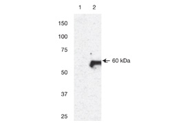
Figure 9. Western blot of 3T3 cell lysates labeled with AKT [pS473] - Recombinant Rabbit Monoclonal Antibody. Rabbit anti-AKT [pS473] (0.1 µg/mL) was used to label AKT [pS473] in untreated 3T3 lysates (lane 1) or PDGF-treated 3T3 lysates (lane 2).

Need antibodies in bulk quantities or custom packaging?
Inquire now. Complete Request Form >

Want us to produce a custom antibody for you?
Learn more. Custom Antibody Services >
Resources
For Research Use Only. Not for use in diagnostic procedures.