ABfinity SNAP25 and VAMP1 Antibodies for Studying Neuronal Synaptic Vesicle Exocytosis

Introduction
Download the PDF

The functional unit of the nervous system, the terminally differentiated neuron, is an exquisitely specialized cell with an extraordinary structure that displays a high degree of polarity. From the neuronal cell body arise branching dendrites that receive signals from other neurons and an extended axon that transmits signals to other cells [1]. The functional connections between neurons are governed by specialized structures known as synapses, where membrane-bound synaptic vesicles carry and release neurotransmitters. Hundreds of proteins regulate the proper release of neurotransmitters from these synaptic vesicles. Synaptic plasticity is involved in memory and learning, and improper synaptic function is implicated in a myriad of disorders, including autism, schizophrenia, Alzheimer’s disease, and numerous other neuropsychiatric disorders [2]. Elucidating the biochemical basis of synaptic diversity, composition, and function are therefore fundamental to our understanding and treatment of these disorders.

Soluble NSF attachment protein receptor (SNARE) proteins belong to a multi-member protein family that plays an important role in the release of neurotransmitters through synaptic vesicle fusion and exocytosis.

A synaptosomal-associated protein of 25 kDa (SNAP25) and vesicle-associated membrane protein-1 (VAMP1), also known as synaptobrevin (Syb), are the two major SNARE proteins that mediate synaptic vesicle exocytosis [3,4]. The therapeutic relevance of these proteins is underscored by the effects of certain mutations in disease outcomes. A dominant negative mutation in SNAP25 leads to impaired vesicle trafficking in a mouse model [5], and other mutations are a novel cause of epilepsy and genetic disability [6]. VAMP1 mutations have been reported in congenital myasthenic syndrome and hereditary spastic ataxia [7,8].

In neurons, SNAP25 primarily resides in axons and nerve endings, but also displays significant localization in the cytoplasm, and in the plasma membrane through its palmitoylated cysteine residues [9]. Treatment with nerve growth factor (NGF) enhances expression of SNAP25 1.8–3 fold in PC12 cells [10]. VAMP1 is an integral membrane protein present in synaptic vesicles. VAMP1 and synaptic protein syntaxin interact with SNAP25 after the latter forms a binary complex with another protein, tagmin. This interaction plays a critical role in fusion of the synaptic vesicle to the presynaptic membrane [11].

Results and discussion

The Invitrogen ABfinity Anti-SNAP25 Recombinant Rabbit Monoclonal Antibody and ABfinity Anti-VAMP1 Recombinant Rabbit Oligoclonal Antibody are functional in immunofluorescence and western blotting. Using relevant biological systems and genetic modifications as described below, we establish their high specificity for their targets.

In mouse hippocampal neurons, the ABfinity anti-SNAP25 antibody detects SNAP25 that is enriched in the neuronal processes (Figure 1).

Figure 1. Expression of SNAP25 in primary mouse hippocampal neurons by immunofluorescence using ABfinity SNAP25 monoclonal antibody. (A) SNAP25 expression, (B) DAPI signal, (C) Composite image showing the overlay of SNAP25 and DAPI signals.

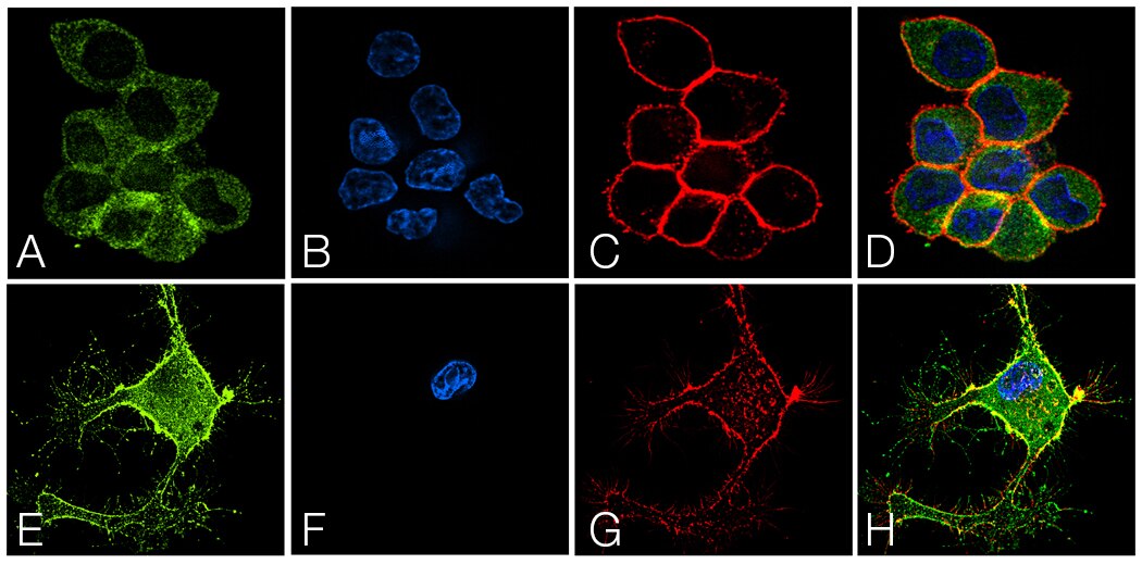
SNAP25 is detected throughout the cytoplasm in undifferentiated PC12 cells; however, upon NGF-induced differentiation, SNAP25 is detected predominantly in the membrane and neuronal processes (Figure 2). These results align with published data about the function and localization of SNAP25 and establishes that the ABfinity anti-SNAP25 antibody specifically detects SNAP25.

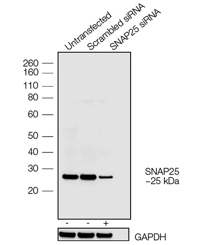
In western blot imaging, the ABfinity anti-SNAP25 antibody detected a single band at the expected molecular weight in PC12 cells. To further confirm the specificity of this antibody, SNAP25 was knocked down by siRNA in PC12 cells and its expression was detected by western blot (Figure 3). Reduced expression of SNAP25 was detected in cell lysates transfected with siRNA compared to untransfected and scrambled controls, confirming that the observed band indeed corresponds to SNAP25.

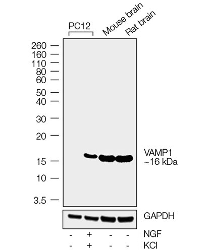
The ABfinity anti-VAMP1 antibody detects a single band by western blot, corresponding to the expected molecular weight of VAMP1 in PC12 cells, and in rat and mouse brain tissue (Figure 4). As expected, VAMP1 is detected in NGF-differentiated PC12 cells but not in undifferentiated cells.

Figure 2. Detection of SNAP25 in PC12 cells by immunofluorescence using ABfinity SNAP25 monoclonal antibody. Panels (A–D) show detection and localization of SNAP25 in the cytoplasm of undifferentiated PC12 cells. Panels (E–H) show increased signal of SNAP25 in the membrane and neuronal processes of NGF-differentiated PC12 cells.

SNAP25 is detected throughout the cytoplasm in undifferentiated PC12 cells; however, upon NGF-induced differentiation, SNAP25 is detected predominantly in the membrane and neuronal processes (Figure 2). These results align with published data about the function and localization of SNAP25 and establishes that the ABfinity anti-SNAP25 antibody specifically detects SNAP25.

In western blot imaging, the ABfinity anti-SNAP25 antibody detected a single band at the expected molecular weight in PC12 cells. To further confirm the specificity of this antibody, SNAP25 was knocked down by siRNA in PC12 cells and its expression was detected by western blot (Figure 3). Reduced expression of SNAP25 was detected in cell lysates transfected with siRNA compared to untransfected and scrambled controls, confirming that the observed band indeed corresponds to SNAP25.

The ABfinity anti-VAMP1 antibody detects a single band by western blot, corresponding to the expected molecular weight of VAMP1 in PC12 cells, and in rat and mouse brain tissue (Figure 4). As expected, VAMP1 is detected in NGF-differentiated PC12 cells but not in undifferentiated cells.

The blots were probed with ABfinity Anti-VAMP1 Recombinant Rabbit Oligoclonal Antibody (2.5 μg/mL) and detected by chemiluminescence using Invitrogen Goat Anti-Rabbit IgG (H+L) Superclonal Secondary Antibody, HRP conjugate (0.4 μg/mL, 1:4,000 dilution). A 16 kDa band corresponding to VAMP1 was observed specifically in the NGF-differentiated PC12 cell line and tissues tested. Known quantities of protein samples were electrophoresed using Invitrogen NuPAGE 10% Bis-Tris Protein Gel, XCell SureLock Mini-Cell and XCell II Blot Module and Sharp Pre-Stained Protein Standard. Resolved proteins were then transferred onto a nitrocellulose membrane with the Invitrogen iBlot Dry Blotting System. The membrane was probed with the relevant primary and secondary antibodies following blocking with 5% skimmed milk. Chemiluminescence detection was performed using Invitrogen Novex ECL Chemiluminescent Substrate Reagent Kit.

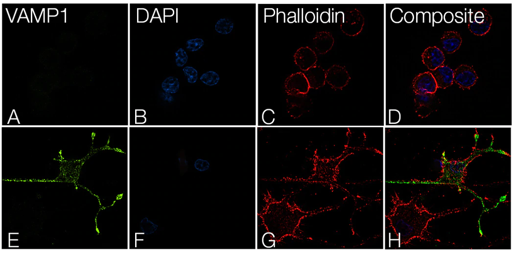
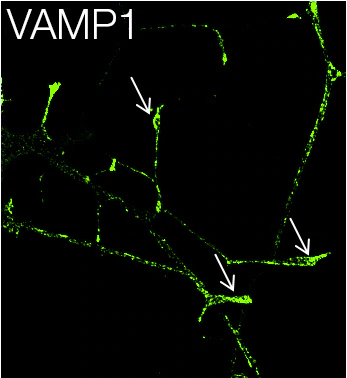
Similarly, there is a strong increase in immunofluorescence signal with the anti-VAMP1 antibody in NGF-differentiated PC12 cells compared to the undifferentiated cells (Figure 5). Studies have reported an accumulation of synaptic vesicles in growth cones [7]. Consistent with this data, immunofluorescence in NGF-differentiated PC12 cells with the anti-VAMP1 antibody also displayed enhanced signal in growth cones (Figure 6), underscoring the specificity of this antibody for its target.

Figure 5. Detection of VAMP1 in PC12 cells by immunofluorescence using ABfinity VAMP1 oligoclonal antibody. Panels (A–D) show no expression of VAMP1 in undifferentiated PC12 cells. Panels (E–H) show increased signal of VAMP1 on neurite tips of NGF-differentiated PC12 cells.

Figure 3. Detection of SNAP25 in PC12 cells upon siRNA transfection using ABfinity SNAP25 monoclonal antibody.


Figure 4. Western blot analysis on whole-cell extracts (30 μg lysate) of PC-12 (lane 1), PC12 treated with NGF and KCl (NGF at 200 nM, KCl at 50 nM for 7 days) (lane 2), and tissue extracts (30 μg lysate) of mouse brain (lane 3) and rat brain (lane 4).

Figure 6. Detection of VAMP1 on growth cones (indicated by arrows) of NGF-differentiated PC12 cells using ABfinity VAMP1 oligoclonal antibody.


Conclusion

Together, the data presented in this application note describe the high specificity of ABfinity recombinant anti-SNAP25 monoclonal antibody and ABfinity anti-VAMP1 oligoclonal antibody to their respective targets, and their performance in applications that are most routinely used to study these proteins. These antibodies should serve as key reagents in the study of neuronal disease by enabling researchers to design and conduct elegant and nuanced experiments in biologically appropriate model systems.